Early Pregnancy Complications No. 2

Learning Outcomes

On completing this case study, reading associated articles and answering related questions, you will;

  • Appreciate the extreme variation in the presentation of molar pregnancy
  • To understand the roles of varying imaging modalities in determining the stage of disease and the chemotherapy score

Case History

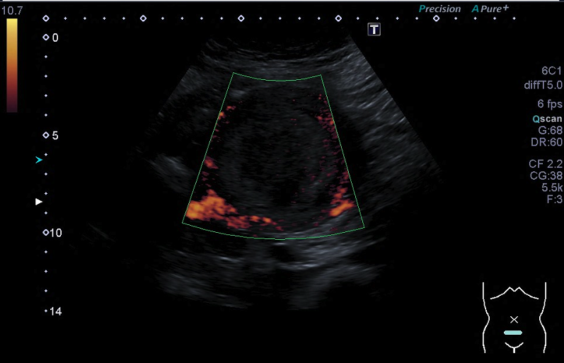
A 31-year-old lady presented to her local accident and emergency department with acute onset abdominal pain. A surgical emergency was excluded but abdominal examination identified a mass arising from the pelvis, which reached the umbilicus. Vaginal examination visualised vesicular tissue protruding from the cervical os that was sent for histological analysis. A serum HCG level was >200,000 and no intrauterine pregnancy was imaged using ultrasound. The working diagnosis was a molar pregnancy but subsequent imaging and histopathology confirmed a diagnosis of metastatic choriocarcinoma.

Diagnosis: Metastatic choriocarcinoma

Management Issues

Molar pregnancies are usually identified early, either because of early symptom investigation, because of the sonographic appearance of early pregnancy scans or following histological analysis of terminations, uterine evacuations and medical / spontaneous miscarriages. These cases rarely present in such an advanced state. Because of the high response rate of the abnormal tissue to chemotherapy the management always consists of the same components – uterine evacuation to remove tumour mass and obtain a tissue sample and either single or multi agent chemo therapy. The critical step in this case was to determine the risk score (see below), which determined the chemotherapy protocol, and this depended on a combination of radiological imaging techniques.

Risk Score0124
Age<40>40--
Antecedent pregnancyMaleAbortionTerm
Months from index pregnancy<44-67-13>13
HCG<10001000-10,00010,000-100,000100,000+
Largest tumour size-3-5cm>5cm
SiteLungSpleen / KidneyGIBrain / Liver
Number-1-45-8>8
Previous chemo--Single agentMulti agent

Discussion

At one end of the spectrum of gestational trophoblastic are the benign conditions of complete and partial moles and at the malignant end are the rare histological types of invasive moles choriocarcinoma and placental site trophoblastic tumour. All respond well to chemotherapy and the cure rates are greater than 98%. Radiological imaging is key in both diagnosis and follow up. Ultrasound is used to exclude a viable pregnancy, determine the largest tumour diameter and asses abnormal blood flow with power Doppler. MRI can determine the extent of the disease in the pelvis and local invasion. It is also the most suitable modality for imaging the brain for evidence metastases. T2 weighted images are best used for CNS imaging as the high intensity signal from CSF and metastases (high water content) contrast best against the surrounding tissue. T1 weighted images are best used for imaging the pelvis where fat appears to have the highest signal intensity. CT is most suited for looking for the presence of extra pelvic disease in the lungs and abdominal organs. No one imaging modality is superior to another; rather they all complement each other.

References

RCOG: Gestational trophoblastic Disease:

http://www.rcog.org.uk/womens-health/clinical-guidance/management-gestational-trophoblastic-neoplasia-green-top-38

Charing Cross Gestational trophoblastic unit:

http://www.hmole-chorio.org.uk

Early pregnancy complications No. 2

You may wish to answer these questions more than once to improve your score. Following that e-mail admin@bsgi.org.uk requesting the correct answers and explanations.

"*" indicates required fields

This field is hidden when viewing the form
What is the approximate quoted incidence of gestational trophoblastic disease?*
How is the definitive diagnosis of molar pregnancy made?*
The chest x-ray is in keeping with which FIGO stage?*
The CRX shows:*
Which of the following can CT not reliably comment on:*
How would CNS mets appear o a T2 weighted image of the brain?*
The first ultrasound image is taken in which plane:*
The second ultrasound image highlights?*

Events

We have many events throughout the year, see what's coming up!

View Events

Events

We have many events throughout the year, see what's coming up!

View Events