Early Pregnancy Complications No. 1

Lorem ipsum dolor sit amet, consectetur adipiscing elit. Ut lobortis nibh sed mauris aliquet maximus. Praesent convallis mauris in ante commodo, quis ultrices urna feugiat. Nam et auctor diam. Vestibulum ut suscipit lectus. Cras feugiat mauris a leo sagittis, ac viverra odio pulvinar.

Mauris dapibus mauris eu turpis hendrerit, in vulputate nunc volutpat. Orci varius natoque penatibus et magnis dis parturient montes, nascetur ridiculus mus.

On completing this case study, reading associated articles and answering related questions, you will;

  • Expand your knowledge on the spectrum of symptoms and signs of ectopic pregnancy presentation,
  • Become familiar with the role of diagnostic tools and in particular the appearance of ectopic pregnancy on ultrasound,
  • Develop a critical approach to evaluating the quality of ultrasound images,
  • Understand the advantages and disadvantages of different treatment options,
  • Be able to reflect and test the acquired knowledge.
Case History

A 36 year old nulliparous lady presents to A+E with worsening abdominal pain. She found earlier that morning she was pregnant (positive urine dip stick). This was an unplanned pregnancy. Later that morning she fainted after having a shower. Her abdominal pain became worse and was now associated with loose stools. There was no nausea or vomiting. Her husband called an ambulance as he was concerned about her condition.

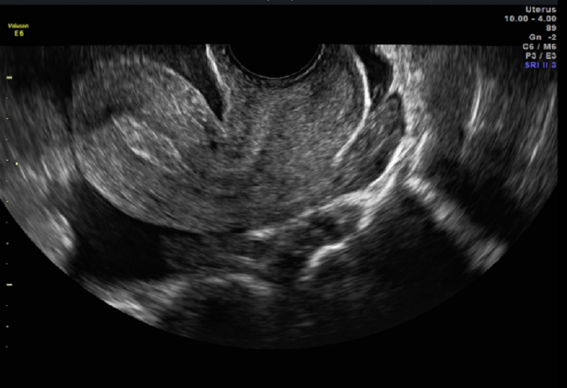
Sagittal view of the uterus showing thickened endometrium with no obvious gestational sac.

On arrival in accident and emergency she was haemodynamically stable. She was well perfused, her abdomen was tender and there was no peritonism. There was no vaginal bleeding and bimanual examination was generally tender. Her Hb was 99g/L and her HCG was 1765IU/L. An ultrasound scan was requested.

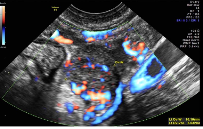
Adnexal mass adjacent to the ovary and showing abnormal vascularity.

Diagnosis: Ectopic pregnancy

Management Issues

In a haemodynamically unstable patient with suspicion of ectopic pregnancy, prompt surgical intervention is the management of choice.

In a stable patient, ultrasound should be the first-line imaging modality. If an intrauterine pregnancy is confirmed, other causes for the presentation should be considered such as corpus luteum accident, pelvic inflammatory disease or other non-gynaecological causes.

Discussion

Ectopic pregnancies may present with no risk factors and only subtle clues in the history. A high index of suspicion is needed so as not to delay the diagnosis. Healthy women compensate haemodynamically, even with significant intra-abdominal bleeding, and maintain their BP but may become unstabe rapidly.

An ultrasound performed in A+E would have significantly raised the index of suspicion of a ruptured ectopic pregnancy (free fluid + positive pregnancy test + pain). Transvaginal ultrasound

is both sensitive and specific for the diagnosis of ectopic pregnancy.

Ultrasound Features in this Case Include

Anteverted uterus with no evidence of an inter-uterine pregnancy (no gestational sac) and a thickened endometrium only

Uterus surrounded by free fluid, both anterior to it and in the pouch of Douglas

Floating loops of bowel in the pelvis

Visible internal iliac vessel adjacent to the ovary partly highlighted with colour flow Doppler in Figure 2.

Adnexal mass adjacent to the ovary showing abnormal vascularity.

Further Reading

Green Top Guideline No. 21, Management of tubal pregnancy. www.rcog.org.uk

The accuracy of transvaginal ultrasound for the diagnosis of ectopic pregnancy prior to surgery. Condous et al. Go to Free article: >> Human Reproduction Vol. 20, No. 5 pp. 1404–1409, 2005

Early pregnancy complications No. 1

You may wish to answer these questions more than once to improve your score. Following that e-mail admin@bsgi.org.uk requesting the correct answers and explanations.

"*" indicates required fields

This field is hidden when viewing the form
Regarding the incidence of ectopic pregnancy, it is:*
Which is the most infrequently seen site of ectopic pregnancy?*
The sensitivity and specificity of TVUSS in diagnosing ectopic pregnancy is:*
Which is not a recognised ultrasound finding associated with ectopic pregnancy?*
What is the most common ultrasound finding associated with ectopic pregnancy?*
What is the best practice advised for the follow up of ectopic pregnancy when managed conservatively?*
Free fluid in the pelvis with a positive pregnancy test, can be associated with:*
The technique applied to the above image (2) is:*
In future pregnancies, best practice is:*

Events

We have many events throughout the year, see what's coming up!

View Events

Events

We have many events throughout the year, see what's coming up!

View Events